What is pregnancy screening? How many times and at what period is a pregnant woman screened? What is screening and how is it done? What is CTE during pregnancy on ultrasound
Prenatal screenings cause a lot of conflicting opinions and reviews. Some are convinced of their necessity, others are confident of their complete inexpediency. What are these tests, and should all pregnant women really undergo them? We decided to look into this issue.
Prenatal screening is a complex of studies, the main goal of which is to identify a risk group of pregnant women with possible malformations of the child (such as: Edwards syndrome, neural tube defects (anencephaly), Cornelia de Lange syndrome, Smith Lemli Opitz syndrome, triploidy, Patau syndrome ).
Despite the fact that screenings include two fairly proven diagnostic methods - and ultrasound, their reliability and safety still cause a lot of controversy.
Cons No. 1: Ultrasound is harmful to the baby
There is a fairly widespread opinion that ultrasound negatively affects the child’s nervous system and irritates him - during the examination, children often try to hide from the machine and cover their heads with their hands. Therefore, children whose mothers regularly had ultrasound scans during pregnancy are more restless compared to babies whose mothers refused ultrasound diagnostics. Is it really?
According to doctors, ultrasound cannot cause any harm to the baby - modern equipment is absolutely safe. Therefore, official medicine insists that absolutely all pregnant women undergo an ultrasound. After all, timely diagnosis allows, firstly, to see the full picture of the course of pregnancy, and secondly, if necessary, to correct certain problems.
An ultrasound examination is carried out at least three times during pregnancy (on, on, and on), but if necessary, the doctor may recommend having it more often.
The data obtained from the ultrasound of the first prenatal screening (on) are considered especially important. At this time during the study:
- the number of embryos in the uterus and their viability are determined;
- more accurate is set;
- gross malformations are excluded;
- the thickness of the collar space is determined - TVP (i.e. the amount of subcutaneous fluid on the back surface of the child’s neck is measured - normally TVP should not exceed 2.7 mm);
- The presence or absence of the nasal bone is examined.
For example, in children with Down syndrome, the fluid content is much higher than normal, and the nasal bone is often not visualized.
Cons No. 2: biochemical blood test gives unreliable screening result
Many mothers are sure that it is impossible to draw any reliable conclusions from one analysis - too many factors can affect the result. And they are partly right. However, you need to take a closer look at the analysis process in order to understand on what basis the doctor makes his conclusion.
A biochemical analysis is carried out to determine the level of specific placental proteins in the blood. During the first screening, a “double test” is done (that is, the level of two proteins is determined):
- PAPPA (pregnancy associated plasma protein or pregnancy-associated plasma protein A);
- free beta subunit (human chorionic gonadotropin).
Changes in the levels of these proteins indicate the risk of various chromosomal and some non-chromosomal disorders. However, identifying an increased risk does not mean that there is something wrong with the baby. Such indicators are only a reason for more careful monitoring of the course of pregnancy and the development of the child. As a rule, if the first trimester screening results in an increased risk for any indicators, the expectant mother is asked to wait for the second screening. In case of serious deviations from the norm, the woman is referred for consultation to a geneticist.

The second screening takes place on. This study includes a "triple" or "quadruple" test. Everything happens the same as in the first trimester - the woman takes a blood test again. Only in this case, the results of the analysis are used to determine not two, but three (or, accordingly, four) indicators:
- free beta subunit of hCG;
- alpha-fetoprotein;
- free estriol;
- in the case of a quadruple test, also inhibin A.
As in the first screening, the interpretation of the results of the 2nd screening is based on the deviation of the indicators from the average statistical norm according to certain criteria. All calculations are carried out using a special computer program, after which they are carefully analyzed by a doctor. In addition, when analyzing the results, many individual parameters are taken into account (race, presence of chronic diseases, number of fetuses, body weight, bad habits, etc.), since these factors can influence the value of the studied indicators.
In order to obtain the most reliable results, the data from studies of the first and second trimester must be correlated together.
If, as a result of the research, any abnormalities in the development of the fetus are revealed, the woman may be offered to undergo repeated screening or immediately referred to a geneticist for consultation. If necessary, he may prescribe additional tests to make a more accurate diagnosis (for example, amniotic fluid testing, chorionic villus biopsy). However, due to the fact that these studies are not entirely safe and can cause various complications during pregnancy (risk, development of group or infection of the fetus, etc.), they are prescribed only in case of a high risk of pathology. However, such complications do not occur so often - in 12% of cases. And, of course, all research is carried out only with the consent of the expectant mother.
Thus, the first two arguments against, from the point of view of scientific medicine, are not convincing, and rather they should be reformulated as follows: prenatal screenings are safe for the expectant mother and her baby, and all conclusions are made by the doctor taking into account a whole range of individual factors.
“Con” No. 3: “I have good heredity - I don’t need screenings”
Some mothers do not see the point of undergoing screenings during pregnancy - all relatives are healthy, what problems could there be? Indeed, there are certain groups of women who are first recommended to undergo testing to identify possible pathologies in the development of the child. These are women over 35-40 years old (since after this age the risk of developing abnormalities in a child increases several times) and expectant mothers with certain diseases (for example, diabetes). Of course, those mothers whose families already have children or relatives with genetic diseases are also at risk. However, the majority of doctors (not only in Russia, but also in many European countries and America) are of the opinion that all women must undergo prenatal screening, especially if it is their first pregnancy.
“Con” No. 4: “I’m afraid of hearing a bad diagnosis”
This is perhaps one of the strongest arguments against screening during pregnancy. Expectant mothers are very frightened by the possibility of hearing something bad about the baby’s development. In addition, medical errors are also a concern - sometimes screenings give false positive or false negative results. There are cases when the mother was told that the child was suspected of having Down syndrome, and subsequently a healthy baby was born. Of course, needless to say, such news greatly affects the emotional state of the mother. After a preliminary conclusion is made, a woman spends the rest of her pregnancy in constant worry, but this is also not at all beneficial for the baby’s health.
However, we should not forget that the results of prenatal screenings in no way serve as a basis for making a diagnosis. They only identify probable risks. Therefore, even a positive screening result will not be a “sentence” for the child. This is just a reason to get professional advice from a geneticist.

“Cons” No. 5: identified potential deviations in the development of the child cannot be corrected
This is true - there is no way to cure or correct chromosomal disorders. Therefore, impressionable and vulnerable mothers, as well as women who are determined to maintain their existing pregnancy under any circumstances, may only receive another reason for worry as a result of the screenings they have undergone. Perhaps, indeed, the best way out in such a situation would be to refuse research so that the mother can calmly wait for the birth of the baby.
And still...
An undoubted advantage of prenatal screenings is the opportunity to obtain information about the development of the child at a fairly early stage of pregnancy, go for a consultation with a geneticist, and undergo, if necessary, all additional examinations. After all, having the data, the expectant mother can quite consciously make a decision on the further development or termination of pregnancy.
The most important argument against: poor health of the expectant mother at the time of the study
Any, even slight increase in body temperature, colds (acute respiratory infections, acute respiratory viral infections), any other viral and infectious diseases, and even stress are a clear contraindication for screening. After all, each of these factors can distort analysis data. That is why, before going to donate blood, the expectant mother must undergo an examination by a gynecologist - the doctor will assess her general condition.
Today, prenatal screenings are not strictly mandatory, but most doctors are confident in the need for these studies. The right to make a decision remains with the pregnant woman, so after weighing all the pros and cons, each woman will make a choice - for some it is important to control the situation and receive all possible information as early as possible, while for others it is much calmer to make do with only the mandatory minimum examinations, just enjoy your pregnancy and believe in the best.
Screening - translated from English, this word means sorting or selection. In short, perinatal screening is a special set of tests, tests, and studies that can give a clear idea of possible deviations in the development of the unborn child.
All screening is divided into the number of trimesters, since during each period of gestation the expectant mother is required to undergo the planned tests.
Screenings are divided into double, triple and quarter tests, demonstrating certain hormonal abnormalities during all periods of pregnancy.
The main goal of screening is to separate risk categories for the development of congenital defects in the fetus: Down syndrome, Edwards syndrome, neural tube defects. Based on the indicators of the ultrasound examination and the results of a blood test taken from a vein, the result is calculated.
Naturally, when processing information, the woman’s personal information is taken into account (from age, weight, bad habits to the use of hormonal drugs during pregnancy).
What screening tests should be taken during pregnancy?
An ultrasound should examine the thickness of the nuchal translucency (nuchal translucency). Its coefficient, if it exceeds 2-2.5 cm, indicates the possible presence of Down syndrome in the child.TVP is measured at strictly limited periods of pregnancy - from 11 to 14 weeks, more precisely - up to 12 weeks. Later, the fetus will grow up and the TVP indicators will lose their information content.
In the first trimester, blood is donated for the hormones b-hCG and PAPP-A.
The second screening (16-18 weeks) does not include an ultrasound scan - the indications for it are taken from the first. And blood must be donated for the hormone b-hCG, the protein Alpha protein AFP and estriol - that is, the so-called “triple test”.
Screening test results
You must wait about three weeks for results. Analysis indicators are expressed not in numbers, but in MoM, which means multiplicity in medicine. The median is the statistical average for the given marker. According to the norm, MoM should be within the range of 0.5-2.0. If, based on the tests, a deviation from the norm is revealed, it means that there is some pathology in the development of the fetus. Elevated hCG may indicate the following abnormalities: chromosomal developmental defects, multiple births, Rh conflict. Reduced hCG indicates an ectopic pregnancy, threatened miscarriage, or undeveloped pregnancy. An increase or decrease in AFP indicates probable chromosomal abnormalities.
The sum and combinations of deviations in the proportions of hormones can also indicate the presence of pathologies. Let’s say that in Down syndrome, the AFP indicator is underestimated, and hCG, on the contrary, is overestimated. The hallmark of an unclosed neural tube is increased levels of alpha protein (AFP) and decreased levels of the hormone human chorionic gonadotropin (hCG). In Edwards syndrome, the test hormones are reduced.
If there is a high risk
If the risk is high, the woman is referred for consultation to a genetic specialist. Here you need to make a very important decision in life. The malformations indicated by your measurements cannot be treated. Here you will be given information that you will most likely have a “different” child.The geneticist will study your indicators, information about your pedigree, clarify whether hormonal treatment was used to maintain the pregnancy (Utrozhestan, Duphaston) and will certainly warn that there is no way to find out with one hundred percent accuracy whether the baby has pathologies, except by invasive methods. These methods are not very harmless: chorionic villus biopsy, amniocentesis (taking amniotic fluid through a puncture in the abdomen), cordocentesis (puncture from the fetal umbilical cord). There is a certain risk in conducting invasive research.
Unfortunately, today screenings provide little information. The unreliability and fallibility of non-invasive studies is quite high. Some doctors even argue about the advisability of such procedures.
In the first three months of pregnancy, absolutely all women undergo this painless procedure.Provides the opportunity to recognize pathologies in fetal development. It consists of an ultrasound examination and blood tests. To carry out diagnostics, all personal data of a woman are taken into account (from age, weight, presence of chronic diseases to bad habits). Blood is taken from her vein and an ultrasound is performed.
Timing of the first screening during pregnancy
All these actions are carried out at 10-13 weeks of gestation. Despite such a short period, they help identify genetic and chromosomal abnormalities in the fetus.All conclusions about the development of the unborn child are made based on the results of a sum of research and analyses. If the diagnosis determines a high probability of abnormalities in the formation of the baby, the woman is sent for amniocentosis and IVS.
Risk group:
- Women who are whiter than 35 years old.
- Those expectant mothers who had children with Down syndrome or other genetic abnormalities.
- Pregnant women who have already given birth to children with disabilities or who have had miscarriages in the past.
Stage of preparation for the first screening
Preparation for the first screening takes place in the antenatal clinic under the guidance of a gynecologist.- Try to conduct the blood test and ultrasound on the same day and in the same laboratory.
- Do a blood test on an empty stomach and abstain from sexual intercourse to eliminate the possibility of distorting the results.
- Weigh yourself before going to the clinic - this is necessary to fill out the form.
- Before the procedure, you should not drink water, at least not more than 100 ml.
How does the first screening process work?
First stage– biochemical. This is the process of blood testing. Its task is to identify such abnormalities as Down syndrome, Edwards syndrome, and defects in the formation of the brain and spinal cord in the fetus.The results of a blood test during the first screening do not provide reliable data for making a diagnosis, but give rise to additional research.
Second phase- This is an ultrasound scan of the first three months of pregnancy. It determines the development of internal organs and the location of the limbs. In addition, measurements of the child’s body are taken and compared with age-appropriate standards. This screening examines the location and structure of the placenta and nasal bone of the fetus. Usually at this stage it is visible in 98% of children.
Norms for first screening during pregnancy
Also, the first screening determines a multiple pregnancy if all indicators exceed the norm.- If the test results are higher than normal, then the risk of Down syndrome in the unborn child is high. If they are below normal, then Edwards syndrome is possible.
- The PAPP-A norm is another coefficient for the first screening during pregnancy. This is plasma protein A, the level of which increases throughout pregnancy, and if this does not happen, then the unborn child has a predisposition to diseases.
- If PAPP-A is below normal, the child has a high risk of developing abnormalities and pathologies. If it is higher than normal, but other research results do not deviate from the norm, then do not worry.
To calculate the indicators, it is necessary to use the MoM coefficient, which indicates deviations from the average. During the calculation process, adjusted values are taken that take into account the characteristics of the female body.
If you have any doubts about the results of the screening, repeat it by taking the same blood tests and ultrasound again in another laboratory. This can be done up to 13 weeks of pregnancy.
 Using screening, risk groups for complications, as well as congenital pathologies in the fetus during pregnancy, are determined.
 Repeated screening is carried out during the second trimester, although weeks 16-17 are considered the most effective.
Timing of the second screening during pregnancy
A secondary comprehensive study is carried out to determine the likelihood of the formation of chromosomal abnormalities in the fetus: at this time their probability is quite high.There are three types of second screening:
- ultrasound (detection of abnormalities using ultrasound),
- biochemical (blood parameters),
- combined, where the first two are used.
is a biochemical study of the blood of the expectant mother using certain tests.
More precisely, according to the so-called “triple test”, which studies the level of proteins and hormones, such as: human chorionic gonadotropin (hCG) in the blood, alpha-fetoprotein (AFP), free estyrol. The test becomes “quadruple” when this secondary set of studies also involves taking blood to determine the level of inhibin A.
Studying the concentrations of these hormones and proteins in the blood makes it possible to judge with a high degree of probability the possibility of a child developing Down syndrome, Edwards syndrome, and neural tube defects.
The conclusions of a repeated set of studies may be an indirect indicator of the defective state of the child’s formation and exacerbations of the course of pregnancy. For example, an abnormal level of hCG indicates abnormalities in chromosomes, the danger of the formation of preeclampsia, or the presence of diabetes mellitus in the expectant mother.
Reduced hCG levels may indicate disturbances in the development of the placenta.
Increased or decreased AFP and inhibin A in the blood serum of a pregnant woman is a sign of a disorder in the natural formation of the baby and possible congenital anomalies - open neural tube defects, possibly Down syndrome or Edwards syndrome. If alpha-fetoprotein increases sharply, the fetus may die. If the level of the female steroid hormone - free estriol - changes, the activity of the fetoplacental system can be disrupted: its deficiency suggests probable abnormal functioning of the child.
If the results of a repeated set of studies turned out to be unfavorable, you should not worry ahead of time. They only talk about the estimated risks of deviations, they do not constitute a final diagnosis. In the case where at least a single component of the secondary screening does not fit into the norm, it is necessary to carry out additional research. The indicators of a screening study may be influenced by several reasons: in vitro fertilization, a woman’s weight, the presence of diabetes mellitus, bad habits, such as smoking.
Video about screening
It is easier to prevent any disease than to treat it. Therefore, it is important to know which organs are at risk, whether everything is fine with health, what prevention is needed so that the disease does not strike an unexpected blow. Forewarned is forearmed. This old truth couldn't be more true to the essence of screening.
What is screening
Many people believe that screening and ultrasound (ultrasound) are the same thing. In fact, ultrasound is part of screening.
Any patient can undergo an ultrasound examination if they wish. Screening is prescribed by a doctor, as it includes various tests and tests. This procedure gives a complete picture of a person’s condition and helps not only to identify existing pathologies, but also to identify “weak spots” in order to prevent the occurrence of a particular disease.
What kind of screening is there?
Currently, the most common and recommended by doctors are the following types of screening:
- mammary gland. At the age of 18 to 40 years old, it is enough to do an ultrasound, and then you need to do a mammogram. For example, in England, every woman over 40 years old receives an invitation to a free mammogram once every 3 years, since the risk of breast cancer is very high. According to statistics, every hour one woman in the world dies from this disease. But this is one of the types of cancer that, if diagnosed early, can be treated;
- prostate gland. This is one of the most common cancers in men after 40 years of age. It is known how men do not like any medical procedures, even something as harmless as screening. Therefore, doctors joke that the best way to get a man to undergo testing is to tell their wives about the benefits of the study in the antenatal clinic. A woman can convince her husband better than any medical leaflets;
- cervix- another common threat to women's health. Doctors recommend cervical screening annually after becoming sexually active. This is necessary because cervical cancer develops asymptomatically for a long time and does not cause concern;
- colon or colonoscopy. After 50 years, it is recommended to undergo this procedure for both women and men. Colonoscopy can help detect or prevent colon cancer, which is one of the most common cancers. During the screening process, a biopsy is taken - a piece of tissue, which helps to make the correct diagnosis;
- lungs. The risk group for lung cancer includes smokers, people with chronic bronchitis, working in hazardous conditions - asbestos dust, gasoline fumes, coal dust in mines. The risk of lung cancer increases with age, so regular lung screening is advisable;
- thyroid gland. The thyroid gland regulates metabolism. Any malfunction of this organ can lead to deterioration in health. The threat of thyroid cancer can be detected using ultrasound and medical tests. This simple procedure can prevent the onset of a serious disease or detect it at an early stage, since external signs of the tumor do not appear for a long time;
- stomach or gastroscopy. Gastric cancer is one of the cancer diseases with the highest mortality rate. Annual gastroscopy reduces the risk of the disease. During the procedure, a biopsy is taken. During the examination, the doctor checks not only the stomach, but the esophagus and duodenum, which are often at risk.
Screening and pregnancy
The importance of screening during pregnancy cannot be overestimated. Ideally, every pregnant woman should have this procedure done. Screening allows you to determine the general health of the mother and fetus, identify abnormalities and diseases, and prescribe timely treatment.
During pregnancy, routine ultrasounds are performed. If according to their results there are no pathologies, then screening can be done at the request of the expectant mother. But there are circumstances in which a full examination is necessary. These include:
- age. If a woman is over 35 years old and this is her first birth, the risk of complications is high;
- genetic pathologies. If close relatives of the future parents had hereditary genetic diseases - Down syndrome, microcephaly, hydrocephalus, hemophilia;
- failed pregnancies– miscarriages, premature births, ectopic pregnancy;
- infectious diseases suffered by a woman in early pregnancy;
- medicines, which a woman should take and which can harm the unborn child;
- risk factors– harmful working conditions for the expectant mother, smoking, alcoholism.
Conventionally, the pregnancy period is divided into 3 trimesters, during which prenatal screening is carried out - screening in the womb.
First trimester – up to 12 weeks
Usually, screening is not carried out until the eleventh week, since many signs are simply not expressed yet and cannot be analyzed. At 12-13 weeks, the first screening is done, which includes an ultrasound and a blood test from a vein. Sometimes the doctor asks for a urine test.
An ultrasound examination allows you to determine the exact duration of pregnancy, the main parameters of the child’s body, the volume of amniotic fluid, and the condition of the uterus. The blood undergoes a full biochemical analysis, which makes it possible to determine whether there are any pathologies in the mother and child.
Around the 12th week, the first genetic screening is performed according to indications. It is useful to make an appointment with a genetic specialist. Before the visit, you need to make a complete list of genetic pathologies that occurred in the family of both parents. This will help the doctor determine the risk of similar ailments in the unborn baby. In the future, the doctor will pay attention to the presence of signs of these particular diseases.
The results of tests and studies are analyzed, compared with norms, and the doctor issues a conclusion about the course of pregnancy. By the 12th week, you can already determine the gender of the unborn baby with a 50% probability.
Second trimester – up to 28 weeks
At this time, an ultrasound and blood test are performed again. The doctor determines the size and position of the fetus, the condition of the bones and the location of the umbilical cord. At the same time, a second genetic test is performed. By the 12th week you can almost always determine who will appear - a girl or a boy,
Third trimester – until the 43rd week
Based on the ultrasound, the doctor decides whether a cesarean section is necessary or whether the birth process is normal. More and more women are expressing a desire to resort to caesarean section instead of natural childbirth without medical indications. The reasoning is simple: why bother and suffer - you fell asleep, woke up and received a ready-made baby. The operation will take half an hour, but natural childbirth can take a long time. But doctors are still in favor of a normal birth. Nature is wise, and it is not in vain that all living things are born naturally.
Neonatal screening
Childbirth is over. Your baby has been born. At this time, our biggest concern is his health. Therefore, the World Health Organization recommends neonatal or newborn screening for all newborn children. This makes it possible to diagnose congenital genetic diseases long before they make themselves felt.
Blood is taken from the child's heel approximately on the 4th day of his life. Results are received in 2 weeks. If there are deviations from the norm, a detailed examination of the child’s health condition is prescribed.
The screening procedure itself does not cause any discomfort. Ultrasound is one of the few pleasant medical procedures when a special probe is passed over the patient over the area being examined, and on the monitor you can see what is inside you. It is especially touching for the expectant mother to see the baby.
If you have been scheduled for screening, there is no reason to worry. Do not neglect the procedure, because it can save the life of you and your loved ones.
Screening - what it is, types and why it is important to do during pregnancy
It is easier to prevent any disease than to treat it. Therefore, it is important to know which organs are at risk, whether everything is fine with health, what prevention is needed so that the disease does not strike an unexpected blow. Forewarned is forearmed. This old truth couldn't be more true to the essence of screening.
What is screening
Many people believe that screening and ultrasound (ultrasound) are the same thing. In fact, ultrasound is part of screening.
Any patient can undergo an ultrasound examination if they wish. Screening is prescribed by a doctor, as it includes various tests and tests. This procedure gives a complete picture of a person’s condition and helps not only to identify existing pathologies, but also to identify “weak spots” in order to prevent the occurrence of a particular disease.
What kind of screening is there?
Currently, the most common and recommended by doctors are the following types of screening:
- mammary gland. At the age of 18 to 40 years old, it is enough to do an ultrasound, and then you need to do a mammogram. For example, in England, every woman over 40 years old receives an invitation to a free mammogram once every 3 years, since the risk of breast cancer is very high. According to statistics, every hour one woman in the world dies from this disease. But this is one of the types of cancer that, if diagnosed early, can be treated;
- prostate gland. This is one of the most common cancers in men after 40 years of age. It is known how men do not like any medical procedures, even something as harmless as screening. Therefore, doctors joke that the best way to get a man to undergo testing is to tell their wives about the benefits of the study in the antenatal clinic. A woman can convince her husband better than any medical leaflets;
- cervix- another common threat to women's health. Doctors recommend cervical screening annually after becoming sexually active. This is necessary because cervical cancer develops asymptomatically for a long time and does not cause concern;
- colon or colonoscopy. After 50 years, it is recommended to undergo this procedure for both women and men. Colonoscopy can help detect or prevent colon cancer, which is one of the most common cancers. During the screening process, a biopsy is taken - a piece of tissue, which helps to make the correct diagnosis;
- lungs. The risk group for lung cancer includes smokers, people with chronic bronchitis, working in hazardous conditions - asbestos dust, gasoline fumes, coal dust in mines. The risk of lung cancer increases with age, so regular lung screening is advisable;
- thyroid gland. The thyroid gland regulates metabolism. Any malfunction of this organ can lead to deterioration in health. The threat of thyroid cancer can be detected using ultrasound and medical tests. This simple procedure can prevent the onset of a serious disease or detect it at an early stage, since external signs of the tumor do not appear for a long time;
- stomach or gastroscopy. Gastric cancer is one of the cancer diseases with the highest mortality rate. Annual gastroscopy reduces the risk of the disease. During the procedure, a biopsy is taken. During the examination, the doctor checks not only the stomach, but the esophagus and duodenum, which are often at risk.
Screening and pregnancy
The importance of screening during pregnancy cannot be overestimated. Ideally, every pregnant woman should have this procedure done. Screening allows you to determine the general health of the mother and fetus, identify abnormalities and diseases, and prescribe timely treatment.
During pregnancy, routine ultrasounds are performed. If according to their results there are no pathologies, then screening can be done at the request of the expectant mother. But there are circumstances in which a full examination is necessary. These include:
- age. If a woman is over 35 years old and this is her first birth, the risk of complications is high;
- genetic pathologies. If close relatives of the future parents had hereditary genetic diseases - Down syndrome, microcephaly, hydrocephalus, hemophilia;
- failed pregnancies– miscarriages, premature births, ectopic pregnancy;
- infectious diseases suffered by a woman in early pregnancy;
- medicines, which a woman should take and which can harm the unborn child;
- risk factors– harmful working conditions for the expectant mother, smoking, alcoholism.
Conventionally, the pregnancy period is divided into 3 trimesters, during which prenatal screening is carried out - screening in the womb.
First trimester – up to 12 weeks
Usually, screening is not carried out until the eleventh week, since many signs are simply not expressed yet and cannot be analyzed. At 12-13 weeks, the first screening is done, which includes an ultrasound and a blood test from a vein. Sometimes the doctor asks for a urine test.
An ultrasound examination allows you to determine the exact duration of pregnancy, the main parameters of the child’s body, the volume of amniotic fluid, and the condition of the uterus. The blood undergoes a full biochemical analysis, which makes it possible to determine whether there are any pathologies in the mother and child.
Around the 12th week, the first genetic screening is performed according to indications. It is useful to make an appointment with a genetic specialist.
Before the visit, you need to make a complete list of genetic pathologies that occurred in the family of both parents. This will help the doctor determine the risk of similar ailments in the unborn baby.
In the future, the doctor will pay attention to the presence of signs of these particular diseases.
The results of tests and studies are analyzed, compared with norms, and the doctor issues a conclusion about the course of pregnancy. By the 12th week, you can already determine the gender of the unborn baby with a 50% probability.
Second trimester – up to 28 weeks
At this time, an ultrasound and blood test are performed again. The doctor determines the size and position of the fetus, the condition of the bones and the location of the umbilical cord. At the same time, a second genetic test is performed. By the 12th week you can almost always determine who will appear - a girl or a boy,
Third trimester – until the 43rd week
Based on the ultrasound, the doctor decides whether a cesarean section is necessary or whether the birth process is normal. More and more women are expressing a desire to resort to caesarean section instead of natural childbirth without medical indications.
The reasoning is simple: why bother and suffer - you fell asleep, woke up and received a ready-made baby. The operation will take half an hour, but natural childbirth can take a long time. But doctors are still in favor of a normal birth.
Nature is wise, and it is not in vain that all living things are born naturally.
Neonatal screening
Childbirth is over. Your baby has been born. At this time, our biggest concern is his health. Therefore, the World Health Organization recommends neonatal or newborn screening for all newborn children. This makes it possible to diagnose congenital genetic diseases long before they make themselves felt.
Blood is taken from the child's heel approximately on the 4th day of his life. Results are received in 2 weeks. If there are deviations from the norm, a detailed examination of the child’s health condition is prescribed.
The screening procedure itself does not cause any discomfort. Ultrasound is one of the few pleasant medical procedures when a special probe is passed over the patient over the area being examined, and on the monitor you can see what is inside you. It is especially touching for the expectant mother to see the baby.
If you have been scheduled for screening, there is no reason to worry. Do not neglect the procedure, because it can save the life of you and your loved ones.
Source: https://pipla.ru/10173-screening/
Second trimester screening - when and how it is done, what it shows

The screening study is very informative, therefore it is actively used in the practice of managing expectant mothers. It allows you to identify women at risk for complicated pregnancy and detect developmental abnormalities in the fetus.
The first screening test for the expectant mother can be prescribed between 11 and 14 obstetric weeks, the second - from 16 to 20. Ideally, second trimester screening should be carried out at 16-17 obstetric weeks.
Examination of the expectant mother
What is the purpose of the second screening during pregnancy?
Mothers who have already undergone the first comprehensive examination understand perfectly why and when they do the 2nd screening during pregnancy. This procedure pursues the following goals:
- identification of fetal malformations that were not available for study at earlier stages;
- confirmation or refutation of diagnoses made in the first trimester;
- establishing the level of risk of intrauterine pathologies;
- detection of physiological abnormalities in the formation of internal organs and systems of the fetus.
It has been proven that the first screening gives more accurate results than the second. But it cannot serve as a basis for making a final diagnosis. Therefore, if any defects were discovered during the screening of the 1st trimester, the mother is scheduled to undergo a repeat examination. During it, the dynamics of the baby’s development are monitored and appropriate conclusions are drawn.
Ultrasound is an important stage 2 of screening during pregnancy
Who needs to undergo a second screening during pregnancy and why?
Not all pregnant women undergo second trimester screening. It is carried out solely for medical reasons. Among the latest:
- the mother is at risk for bearing a fetus with intrauterine malformations that cannot be treated;
- the age of the pregnant woman is over 35 years;
- there are cases of birth of children with severe developmental defects in the family;
- the baby's father is a close relative of the pregnant woman;
- one of the parents (or both) is a carrier of a genetic disease;
- during the first trimester of pregnancy, the woman suffered an acute bacterial/infectious disease;
- the expectant mother took medications that are contraindicated during pregnancy;
- there is a high risk of miscarriage, the previous birth was difficult, the fetus died.
Assessing the risk of having a child with pathology
It is imperative to undergo screening in the 2nd trimester of pregnancy if a woman is diagnosed with a neoplasm, as well as if the initial comprehensive examination showed a high incidence of giving birth to a child with developmental defects.
Second screening during pregnancy - what geneticists look for
As mentioned above, the second screening during pregnancy is aimed at identifying expectant mothers who are at high risk of developing chromosomal abnormalities in the fetus.
The examination may include:
- ultrasound diagnostics;
- blood tests;
- Ultrasound and simultaneous blood donation.
The last option is called combined. It is preferable because it allows the doctor to obtain the most accurate information.
Submission of laboratory tests by the expectant mother
How is the second screening performed during pregnancy?
A pregnant woman is asked to fill out a questionnaire in which she provides personal information and lists previous illnesses. Afterwards she goes to the laboratory and takes blood tests, then undergoes an ultrasound.
When assessing the results, the doctor must take into account the gestational age. All data is calculated and studied by a special computer program to eliminate the influence of the human factor.
The expectant mother should know that the information obtained after the second screening is not a final diagnosis. If they are disappointing, she will have to undergo additional examination by geneticists. And only after this will it be possible to talk about the baby’s condition and give accurate predictions.
AFP level norms
Blood tests in the second screening during pregnancy
The second screening involves conducting a biochemical examination of the woman’s blood according to tests. Most often, doctors perform a triple test aimed at assessing the levels of:
- human chorionic gonadotropin (hCG);
- free estriol (E3);
- alpha fetoprotein (AFP).
Also, the expectant mother can receive a referral to donate blood to determine the level of inhibin A. Then the laboratory test she completed will no longer be called a triple test, but a quarter test.
It’s good if the mother gets tested between 15 and 16 weeks. Then, if the results are questionable, she will have time to retake them - the latest date for the triple test is the 20th obstetric week.
Alpha-fetoprotein (AFP) level assessment
AFP is a blood plasma protein. First, its production occurs in the yolk sac, and then in the fetal liver. If the level of this compound is low, one can assume Down syndrome or Edwards disease in the baby. In addition, low alpha-fetoprotein indicates:
- diabetes mellitus in women;
- low location of the placenta;
- hypothyroidism.
Hypothyroidism is one of the reasons for decreased AFP levels
An elevated AFP level indicates:
- damage to the fetal nervous system;
- neural tube/abdominal cavity defect;
- abnormal functioning of the child’s kidneys;
- complete or partial absence of the brain in the fetus;
- oligohydramnios;
- Rhesus conflict;
- intrauterine fetal death;
- high risk of miscarriage.
At the same time, a high AFP is considered normal when it comes to multiple pregnancies.
Assessment of human chorionic gonadotropin (hCG) levels
A low level of the hCG hormone can indicate a threat of miscarriage, intrauterine death of a child, non-developing pregnancy, Edwards syndrome.
If the level of human chorionic gonadotropin exceeds the norm, we can talk about severe toxicosis, diabetes mellitus, and the birth of twins.
If the second screening during pregnancy shows that the expectant mother has a high level of hCG, but low levels of AFP and E3, there is a high probability of having a child with Down syndrome.
In multiple pregnancies, high AFP is normal
Determination of free estriol level
The E3 hormone is produced by the placenta and liver of the baby. If pregnancy proceeds without complications, estriol levels gradually increase. Due to this, blood circulation in the vessels of the uterus improves, and the mammary glands are prepared for the process of breastfeeding.
If screening shows that EZ is reduced, the doctor may suggest:
- high risk of miscarriage;
- fetal anemia;
- presence of intrauterine infection;
- delay in the physical development of the baby;
- fetoplacental insufficiency;
- Down syndrome;
- adrenal insufficiency in a child.
It is important to know that the E3 level may decrease due to the use of antibiotics by the expectant mother and fasting.
A high level of EZ is diagnosed when:
- multiple pregnancy;
- high risk of premature birth;
- liver disease.
Blood test for inhibin A
A blood test for inhibin A is rarely included in the second screening during pregnancy. The need to study its level may arise if the triple test showed ambiguous results.
Test for inhibin A during the second screening during pregnancy
Inhibin A levels increase immediately after conception and vary throughout gestation. The hormone is produced by the ovaries if the woman is not pregnant, and by the placenta, the body of the embryo, if conception has taken place.
A low level of inhibin A is observed when there is a threat of miscarriage, a high level is observed when:
- hydatidiform mole;
- fetoplacental insufficiency;
- tumor neoplasms;
- chromosomal abnormalities of the fetus.
Preparing for 2nd trimester screening
It is not at all difficult to physically prepare for the second screening during pregnancy. Need to:
- come to the ultrasound room with a full bladder;
- before taking laboratory tests, do not eat anything; you are allowed to drink only unsweetened water without gas;
- the day before the examination, it is advisable to give up smoked meats and sweets, fatty and fried foods, seafood, cocoa, chocolate, and citrus fruits.
But mentally, preparing for the 2nd trimester screening is very difficult. A woman needs to try to calm down and set herself up for positive results, and not think about anything bad. Even if the data obtained during the study is unsatisfactory, there is no need to sound the alarm ahead of time. Only a consultation with a geneticist will allow everything to fall into place.
It is important for the expectant mother to remain calm even with bad test results
Moreover, no diagnostic examination can give a 100% guarantee of the birth of a sick child. Although rare, screening is wrong. Thus, poor results are the result of diabetes mellitus, multiple pregnancies, obesity, underweight, conceiving a baby through IVF, or incorrectly set due dates. Therefore, you need to believe in a miracle, even if doctors say that it is impossible!
Source: https://babytoday.ru/articles/vtoroy-screening-pri-beremennosti/
What is screening - how to conduct examinations in pregnant women, at what stage and how many times

Screening examinations were introduced as a mandatory procedure for all pregnant women 20 years ago. the task of conducting such analyzes is to identify genetic deviations in the development of the unborn child. The most common include Down syndrome, Edwards syndrome, and a disorder of the formation of the neural tube.
All young mothers should know that screening is a word translated from English as “sifting.” Based on the results of the study, a group of healthy people is determined. Everyone else is prescribed tests that help determine the disease. A comprehensive examination consists of different methods, for example:
- mammography;
- genetic screening;
- computed tomography, etc.
All the technologies described above help in identifying diseases in people who do not yet suspect the presence of pathology.
In some cases, such studies affect the emotional state of a person; not everyone wants to know in advance about the development of a serious illness. This devalues the examination, especially if the patient does not intend to undergo treatment.
In modern medicine, mass examinations are carried out only if there is a real danger to the health of a large number of people.
What is screening for expectant mothers? In this case, a comprehensive examination is necessary to monitor the development of the fetus and assess whether the main indicators correspond to the norm.
When they talk about prenatal screening, we are talking about ultrasound and biochemical examination, for which venous blood is taken. It must be taken on an empty stomach early in the morning so that the substances that came with food do not change the composition.
The screening test will show the level of fetal A-globulin, pregnancy hormone, estriol.
Screening of pregnant women includes an ultrasound examination, which allows you to visually determine abnormalities in the development of the child. An ultrasound shows the main indicators of normal growth - the nasal bone, the collar area. To obtain the most reliable research results, the expectant mother must strictly and clearly follow all the rules of the perinatal examination.
Parents who are concerned about genetics and the risk of developing diseases in their child are interested in how screening is done. At the first stage, the doctor must accurately determine the period.
This affects normal indicators, biochemical blood parameters, and the thickness of the collar space will vary greatly depending on the trimester. For example, TVP at week 11 should be up to 2 mm, and at week 14 – from 2.6 mm.
If the deadline is not set correctly, the ultrasound screening will show an unreliable result. Blood should be donated on the same day as the ultrasound examination so that the data matches.
Biochemical screening
The most accurate way to get an answer to the presence of chromosomal diseases is biochemical screening. Blood is taken on the same day as the ultrasound, early in the morning on an empty stomach.
It is very important to comply with these requirements in order to obtain reliable research results. The sample is taken from a vein, and the material is examined for the presence of a specific substance secreted by the placenta.
The concentration and ratio of marker substances and special proteins in the plasma are also assessed.
It is important to avoid violations during delivery to the laboratory and storage for research. At the reception, the girl will be given a questionnaire containing the following questions:
- about the presence in the family of the child’s father or her people with genetic abnormalities;
- do you already have children, are they healthy;
- whether the expectant mother is diagnosed or not with diabetes;
- whether the woman smokes or not;
- data on height, weight, age.
Screening ultrasound
This is the first stage of examination of the expectant mother. A screening ultrasound is no different from any other ultrasound examination.
During the procedure, the specialist assesses the general condition of the embryo, the rate of development of the child in accordance with the gestational age, the presence of significant developmental defects and any other external abnormalities.
The absence of the latter does not indicate the complete health of the fetus, so a biochemical blood test is performed for a more accurate study.
1st trimester screening
This is the very first screening during pregnancy, which should be carried out at 11-13 weeks of gestation. It is very important to correctly find out the duration of pregnancy in advance. The first appointment necessarily includes an ultrasound examination.
The laboratory sometimes requests ultrasound results to make accurate calculations. A blood test is also performed for protein and hormone: PAPP-A and free b-hCG. This test is called "double".
If a low level of the first is detected, this may indicate:
- The likelihood of developing Down syndrome, Edwards.
- The possibility of developing abnormalities at the chromosomal level.
- The pregnancy stopped developing.
- There is a possibility of having a baby with Cornelia de Lange syndrome.
- There is a risk of miscarriage.
An ultrasound will help the doctor visually assess the course of pregnancy, whether an ectopic conception has occurred, the number of fetuses (if there are more than 1, then he will be able to tell whether there will be fraternal or identical twins).
If the baby’s position is successful, the doctor will be able to assess the fullness of the heartbeat, examine the heart itself, and the mobility of the fetus.
The baby at this stage is completely surrounded by amniotic fluid and can move around like a small fish.
2nd trimester screening
The second screening during pregnancy is carried out between 20-24 weeks. An ultrasound examination and biochemical analysis are ordered again, but this time the test is carried out for 3 hormones.
In addition to checking b-hCG, a check for the level of ACE and estriol is added.
An increased rate of the first indicates an incorrect determination of the term or multiple pregnancy, or may confirm the risk of developing genetic disorders or fetal pathology.
3rd trimester screening
The third pregnancy screening program is carried out between 30-34 weeks.
When doctors do an ultrasound, they evaluate the position of the fetus, the presence or absence of abnormalities in the formation of the baby’s internal organs, whether there is entanglement in the umbilical cord, and whether there are delays in the baby’s development.
During this period, the organs are clearly visible, which helps to accurately determine and provide an opportunity to correct deviations. Experts evaluate the maturity of the placenta, the volume of amniotic fluid, and can tell the gender of the child.
Each stage of pregnancy has certain indicators of the norm of child development and blood composition. Each time the test will show compliance or deviations from standard indicators.
The screening is deciphered by specialists who evaluate the main indicators. Parents are not required to understand the values of TVP or hormone levels in the blood on their own.
The doctor who is monitoring the pregnancy will explain all the indicators in the study results.
Source: https://sovets.net/8414-chto-takoe-screening.html
Screenings during pregnancy

Screening procedures for pregnant women at different stages
The currently popular method of screening during pregnancy causes a lot of controversy among doctors and expectant mothers. A prenatal or prenatal complex of studies allows us to identify threats to pregnancy and the risks of defects in the fetus.
Screenings during pregnancy consist of several types of laboratory and ultrasound diagnostics at different stages, and also determine the risk of defects as a percentage.
After such a comprehensive diagnosis, the results may be indications for detailed invasive examinations and consultations with a geneticist.
However, invasive methods are associated with a high risk for pregnancy and the fetus, and therefore are rarely prescribed.
A high percentage of risk does not mean a guaranteed presence of defects, but requires more in-depth and detailed examinations.
Referring to reviews of screening during pregnancy, we can say that this procedure helps prevent dangers for the mother and child at any stage and during childbirth.
If there are questionable indicators, do not despair; it is necessary to conduct repeated and more in-depth analyzes at the request of specialists.
How is screening done during pregnancy?
The word “screening” translated from English means selection, screening, sorting. This literal definition makes it quite clear that tests and screening tests during pregnancy identify various pathologies. Specific tests included in the mandatory complex can be divided into periods of pregnancy.
Timing of screenings during pregnancy:
- First trimester (11 to 13 weeks).
- Second trimester (16 to 24 weeks).
- Third trimester (based on the complex of previous ones).
Types of screening tests:
- Dopplerometry;
- cardiotocography;
- blood for biochemistry;
- ultrasonography.
Screening during pregnancy by timing
Let us take a closer look at all periods of analysis and the need for each of them to identify pathologies using screening, as well as their normative values and dangerous deviations. Since the tests are not a reasonable cause for stress for expectant mothers, they should be interpreted correctly and, if required, taken again.
First screening during pregnancy
The most important test is considered the first screening in early pregnancy, and even if you feel well, this is not a reason to assume that you do not need a diagnosis. The first screening at the 12th week of pregnancy is carried out by ultrasound and a detailed biochemical blood test. In the latter, accurate indicators of hCG and PAPP-A are required.
During an ultrasound screening during pregnancy at the 12th week, the gynecologist will not only be able to determine the presence of all vital organs and limbs, but also calculate the thickness of the nuchal space (TN).
This special zone between the skin of the neck and the tissues accumulates a kind of liquid, and the threat of pathology depends on its thickness. Often, serious abnormalities indicate pathology or developmental features.
The reliability of the result is ensured by the following conditions:
- the size of the embryo from the coccyx to the crown is usually 44-85 mm;
- sagittal scanning is carried out along the mother’s body, dividing it into two visually equal parts;
- The period for diagnosis should be as close as possible to 12 weeks.
Also, factors such as the quality of equipment and the qualifications of the specialist are of no small importance in this type of diagnosis. The image must be complete and if the fetal head is turned or bent, this may make it somewhat difficult to measure the TVP. The norm for the first screening during pregnancy is considered to be 0.11-0.16 cm, with a maximum value of 0.25 cm.
An increased TVP indicates the presence of chromosomal disorders, for example, Down syndrome or Edwards syndrome and others, which will be revealed during subsequent screenings. However, measuring TVP does not provide complete confidence in the presence of defects, so a blood test is also performed. Also, in rare cases, amniocentesis (amnitic fluid test) and cordocentesis (umbilical cord blood test) may be prescribed.
When testing blood at the first screening, two indicators are taken into account:
- HCG. The level of this hormone secreted by the embryonic membrane is an indicator of pregnancy and usually should not exceed the norm (from 200 thousand to 95 thousand honey/ml).
- The increase indicates the following factors:
- multiple births;
- incorrect diagnostic period;
- diabetes mellitus in a pregnant woman;
- toxicosis;
- chromosomal abnormalities or Down syndrome.
- Low hCG may mean:
- placental insufficiency;
- risk of ectopic pregnancy;
- risk of miscarriage;
- Edwards syndrome.
- RARR-A. Immunomodulatory protein of the placenta should be within normal limits (from 0.33 to 8.55). Its high deviations indicate developmental disorders:
- risk of miscarriage;
- Down, Lange, Edwards syndromes;
- chromosomal disorders.
Second screening during pregnancy
Ultrasound screening during pregnancy in the second trimester allows you to determine the presence of abnormalities in the fetus and the possible risk of miscarriage. The level and possible slowness of development due to the discrepancy between indicators is clarified. The size and location of the embryo relative to the mother's pelvis is determined, on the basis of which the method of delivery is adopted.
An important stage of ultrasound is fetometry, which allows you to measure parts of the body and identify the risk of dysplasia (disorders of skeletal development). Each limb and their compliance are also assessed for the absence of their parts and pathologies. At the stage of fetometry, the following defects are diagnosed in more detail:
- Amniotic fluid. The depth of filling indicates low or high water levels in case of deviations, which leads to defects.
- Face. The nose and jaw are carefully examined to identify clefts, flattened and shortened parts of the face due to Down syndrome, cyclopia or underdeveloped eyes (anophthalmia).
- Brain. Both ventricles, their external and internal formations are measured. If the norm increases by 10 mm, you can be afraid of hydrocephalus (water formations).
- Organs. Diagnosed with the absence of defects and painful formations.
- Placenta. Its location allows you to determine delivery, and its thickness indicates possible infection of the fetus or Rh conflict and developmental delays.
- Spine. Measurement and appearance help identify diseases and hernias.
- Umbilical cord. The number of veins and blood vessels that may indicate hypoxia is assessed.
Biochemical screening during pregnancy up to 20 weeks is usually carried out based on the hormone estriol, hCG and AFP. High deviations from the norm of these indicators may indicate the presence of defects or complications in which the following is observed:
- increased AFP;
- HCG, estriol any.
- Neural tube defects:
- increased AFP;
- HCG and estriol are normal.
- increased AFP, hCG, estriol.
- increased AFP;
- HCG, estriol any.
- increased AFP;
- HCG, estriol any.
- decreased AFP, estriol;
- elevated hCG.
- AFP is normal;
- any estriol;
- hCG decreased.
- decreased AFP, hCG, estriol.
21 pairs of chromosomes:
- decreased AFP, estriol;
- elevated hCG.
13 pairs of chromosomes:
- AFP is normal;
- any estriol;
- hCG decreased.
18 pairs of chromosomes:
- decreased AFP, hCG, estriol.
The hormone alpha-fetoprotein is produced by the liver and gastrointestinal tract of the fetus, which protects it from the effects of the mother's immunity. Its deviations from the norm may indicate:
- an incorrect gestational age previously established may cause differences from the norm;
- Down syndrome is confirmed by the presence of 21 pairs of chromosomes and fully confirms preliminary studies;
- Edwards syndrome is confirmed by 18 pairs of chromosomes and cannot be treated, so a further decision is made to preserve the fetus;
- The death of the child must be confirmed by ultrasound diagnostics, followed by surgical removal of the fetus.
- viral infection can cause necrosis of the fetal liver;
- a hernia or fusion of the abdominal walls is confirmed by ultrasound;
- underdevelopment of the neural tube leads to spina bifida and anencephaly;
- esophageal atresia means the absence of channels that provide the necessary food to the fetus;
- Meckel's syndrome is confirmed by a herniation of the skull or brain at the back of the head on ultrasound.
The hormone estriol is secreted by the placenta and liver of the child and its level normally should gradually increase, since it takes part in the labor readiness of the uterus and mammary glands during lactation:
- liver and urinary tract disease in the mother;
- multiple births;
- The size of the fetus is larger than normal.
- anencephaly;
- hypoplasia;
- infectious diseases of the child;
- early birth;
- Down syndrome;
- Fetal insufficiency of the placenta.
The hCG hormone and its normative deviations, together with indicators of other blood biochemistry tests and ultrasound diagnostics, may indicate possible Down, Patau or Edwards syndromes.
It is also worth taking into account standard deviations of chromosome indicators and previous blood screenings during pregnancy.
Based on a complete detailed diagnosis, the gynecologist can make a diagnosis for the presence or absence of pathologies of fetal development, disturbances in the course of pregnancy and the threat of childbirth.
Third screening during pregnancy
Ultrasound diagnostics at 33 weeks determines:
- General assessment of fetal health.
- Possibility of vices.
- Amniotic fluid and its excess indicate polyhydramnios, which leads to abnormal positioning of the fetus, prolapse of the umbilical cord, bleeding, defects and death of the child.
- Behavioral assessment.
- The placental condition suggests the possibility of a cesarean section if it is located below normal, which can cause bleeding and injury during natural delivery or fetal hypoxia.
- The approximate weight should be about 2.5 kg, since if the weight at 40 weeks exceeds 4 kg, the gynecologist and obstetrician will decide on surgical delivery.
- Dimensions of body parts.
Doppler testing determines the normal functioning of the blood supply between the woman, her placenta and the child, strangulation by the umbilical cord and heart defects. Such screening during pregnancy, if the indicators in the first trimesters are normal, is not prescribed, since the power of diagnostic radiation is higher than with ultrasound. There are several indicators for such a study:
- maternal diseases of the heart and circulatory system, kidneys and liver, diabetes;
- pathologies of fetal development and infectious intraplacental diseases;
- severe pathologies in the anamnesis.
Degrees of defects that Doppler testing reveals:
- Compensated type disorders are common and do not cause serious developmental pathologies. Defects can be corrected with medication and under outpatient supervision. When the condition normalizes, childbirth is resolved naturally, and if it worsens, a decision is made to perform a caesarean section ahead of schedule.
- The subcompensated type of disorder is caused by insufficient blood flow and a reduced amount of oxygen. During outpatient treatment, a course of medications is prescribed and the pregnant woman is under constant medical supervision. If the condition worsens, there is a possibility of early delivery.
- The decompensated type of disorder is quite rare, which requires immediate hospitalization and enhanced medical supervision. The worsened condition implies termination or early delivery.
Doppler echocardiography allows you to examine the child’s cardiac blood flow and detects severe heart pathologies, and also determines the risk of death of the child and allows you to prescribe timely drug treatment. Indications for such a study are:
- accretion of the placenta into the uterus of a pregnant woman;
- slow development and pathologies of the fetus;
- disruption of heart function during ultrasound diagnostics;
- disturbances in fetal mobility and location of the umbilical cord;
- disruption of the circulatory system;
- heartbeat disturbances;
- the need to identify defects and prescribe treatment;
- abnormalities of blood vessels and arteries;
- suspicion of heart pathology;
- defects of internal organs.
Cardiotocography is carried out for all pregnant women and determines the general condition and pulse rate of the fetus, which is carried out only after 32 weeks. During the study, an indicator of the fetal condition is calculated, showing:
- about 1 – absence of defects;
- about 1.5 – probability of violations;
- about 2.5 – risk of developing defect;
- more than 3 – presence of a defect.
Controversial facts about pregnancy screening
There are several common opinions that may influence the decision to conduct such a study:
- A blood test does not allow you to determine the result with high accuracy, but in combination with other methods it gives a more complete picture of the baby’s health and the course of pregnancy.
- The impossibility of correcting defects does exist, but diagnostic indicators make it possible to make an important decision about artificial termination of pregnancy or about the necessary measures during childbirth.
- Poor maternal health is an important aspect for canceling the diagnosis, since general indicators can significantly distort the actual data.
- The fear of a negative diagnosis must be overcome and it is necessary to understand that the diagnosis can only be made by the assumption of pathologies.
- Ultrasound is not harmful when examined with high-quality equipment.
- Good heredity and the absence of diseases in the family do not guarantee protection against fetal pathologies.
- The cost of pregnancy screening is about RUB 1,500, which can only be partially covered by the health center or additional health insurance.
about screening during pregnancy
You can find out how the screening goes and what it shows in the following video.
Some time ago, pregnant women did not even know about such a procedure as prenatal or perinatal . Now all expectant mothers undergo such examination.
What is screening during pregnancy, why is it performed and why are its results so important? Answers to these and other questions that concern many pregnant women about perinatal screening We tried to give in this material.
In order to eliminate any further misunderstanding of the information presented, before moving directly to the consideration of the above topics, it is worth defining some medical terms.
Prenatal screening is a special variation of what is actually a standard procedure, such as screening. Given a comprehensive examination consists of Ultrasound diagnostics and laboratory research, in this particular case biochemistry of maternal serum. Early detection of some genetic abnormalities - this is the main task of such an analysis during pregnancy as screening.
Prenatal or perinatal means prenatal, and by the term screening in medicine, we mean a series of studies of a large segment of the population, which are carried out in order to form a so-called “risk group” susceptible to certain diseases.
Can be universal or selective screening
.
It means that screening studies They are done not only to pregnant women, but also to other categories of people, for example, children of the same age, to establish diseases characteristic of a given period of life.
With help genetic screening Doctors can find out not only about problems in the baby’s development, but also react in time to complications during pregnancy, which a woman may not even suspect.
Often, expectant mothers, having heard that they will have to undergo this procedure several times, begin to panic and worry in advance. However, there is nothing to be afraid of, you just need to ask your gynecologist in advance why you need screening for pregnant women, when and, most importantly, how this procedure is done.
So, let's start with what is standard screening carried out three times during the entire pregnancy, i.e. in every trimester . Let us remind you that trimester is a period of three months.
What it is 1st trimester screening ? First, let's answer a common question about how many weeks it is. first trimester of pregnancy . In gynecology, there are only two ways to reliably determine the due date during pregnancy - calendar and obstetric.
The first is based on the day of conception, and the second depends on menstrual cycle , preceding fertilization . That's why I trimester - this is the period that, according to the calendar method, begins with the first week from conception and ends with the fourteenth week.
According to the second method, I trimester
– this is 12 obstetric weeks. Moreover, in this case, the period is counted from the beginning of the last menstruation. Recently screening
not prescribed to pregnant women. 
However, now many expectant mothers themselves are interested in undergoing such an examination.
In addition, the Ministry of Health strongly recommends that studies be prescribed to all expectant mothers without exception.
True, this is done voluntarily, because no one can force a woman to undergo any kind of analysis.
It is worth noting that there are categories of women who are simply obliged, for one reason or another, to undergo screening, For example:
- pregnant women from thirty-five years of age and beyond;
- expectant mothers whose medical history contains information about the presence of a threat spontaneous ;
- women who in the first trimester had infectious diseases ;
- pregnant women who are forced for health reasons to take medications prohibited for their situation in the early stages;
- women who have had various genetic abnormalities or abnormalities in fetal development ;
- women who have previously given birth to children with any deviations or developmental defects ;
- women who have been diagnosed frozen or regressing pregnancy (cessation of fetal development);
- suffering from narcotic or women;
- pregnant women in whose family or in the family of the father of the unborn child there have been cases of hereditary genetic disorders .
How long does it take to do it? prenatal screening 1st trimester ? For the first screening during pregnancy, the period is set in the interval from 11 weeks to 13 obstetric weeks of pregnancy and 6 days. There is no point in conducting this examination earlier than the specified period, since its results will be uninformative and absolutely useless.
It is no coincidence that a woman’s first ultrasound is performed at 12 weeks of pregnancy. Since this is when it ends embryonic and it begins fetal or fetal period of development of the future person.
This means that the embryo turns into a fetus, i.e. obvious changes occur that indicate the development of a full-fledged living human organism. As we said earlier, screening studies is a set of measures that consists of ultrasound diagnostics and biochemistry of a woman’s blood.
It is important to understand that conducting screening ultrasound in the 1st trimester during pregnancy plays the same important role as laboratory blood tests. Indeed, in order for geneticists to draw the correct conclusions based on the results of the examination, they need to study both the ultrasound results and the biochemistry of the patient’s blood.
We talked about how many weeks the first screening is carried out, now let’s move on to deciphering the results of a comprehensive study. It is really important to take a closer look at the standards established by doctors for the results of the first screening during pregnancy. Of course, only a specialist in this field with the necessary knowledge and, most importantly, experience can give a qualified assessment of the results of the analysis.
We believe that it is advisable for any pregnant woman to know at least general information about the main indicators prenatal screening and their normative values. After all, it is typical for most expectant mothers to be overly suspicious about everything that concerns the health of their future child. Therefore, they will be much more comfortable if they know in advance what to expect from the study.
Interpretation of 1st trimester ultrasound screening, norms and possible deviations
All women know that during pregnancy they will have to undergo more than one ultrasound examination (hereinafter referred to as ultrasound), which helps the doctor monitor the intrauterine development of the unborn child. In order to screening ultrasound gave reliable results, you need to prepare in advance for this procedure.
We are sure that the vast majority of pregnant women know how to do this procedure. However, it is worth repeating that there are two types of research - transvaginal and transabdominal . In the first case, the device sensor is inserted directly into the vagina, and in the second it is in contact with the surface of the anterior abdominal wall.

There are no special preparation rules for transvaginal ultrasound.
If you are undergoing a transabdominal examination, then before the procedure (about 4 hours before the ultrasound) you should not go to the toilet “small”, and it is recommended to drink up to 600 ml of plain water half an hour before.
The thing is that the examination must be carried out on fluid-filled bladder .
In order for the doctor to obtain a reliable result Ultrasound screening, The following conditions must be met:
- the examination period is from 11 to 13 obstetric weeks;
- the position of the fetus should allow the specialist to carry out the necessary manipulations, otherwise the mother will have to “influence” the baby so that he turns over;
- coccyx-parietal size (hereinafter KTR) should not be less than 45 mm.
What is CTE during pregnancy on ultrasound
When performing an ultrasound, a specialist must examine various parameters or sizes of the fetus. This information allows you to determine how well the baby is formed and whether he is developing correctly. The norms for these indicators depend on the stage of pregnancy.
If the value of one or another parameter obtained as a result of ultrasound deviates from the norm upward or downward, then this is considered a signal of the presence of some pathologies. Coccyx-parietal size - This is one of the most important initial indicators of proper intrauterine development of the fetus.
The CTE value is compared with the weight of the fetus and the gestational age. This indicator is determined by measuring the distance from the child’s crown bone to his tailbone. As a general rule, the higher the CTE index, the longer the gestational age.
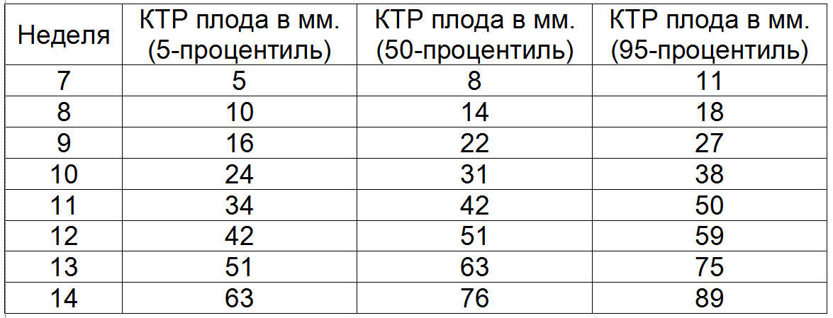
When this indicator is slightly higher or, conversely, slightly lower than the norm, then there is no reason to panic. This only speaks about the developmental characteristics of this particular child.
If the CTE value deviates upward from the standards, then this signals the development of a large-sized fetus, i.e. Presumably, the baby’s weight at birth will exceed the average norm of 3-3.5 kg. In cases where the CTE is significantly less than the standard values, this may be a sign that:
- pregnancy does not develop as expected, in such cases the doctor should carefully check the fetal heartbeat. If he died in the womb, then the woman needs urgent medical attention ( curettage of the uterine cavity ) to prevent possible health hazards ( development of infertility ) and life ( infection, bleeding );
- The pregnant woman's body produces an insufficient amount, as a rule, which can lead to spontaneous miscarriage. In such cases, the doctor prescribes an additional examination for the patient and prescribes medications containing hormones ( , Dufston );
- mother is sick infectious diseases , including sexually transmitted diseases;
- the fetus has genetic abnormalities. In such situations, doctors prescribe additional tests along with, which is part of the first screening test.
It is also worth emphasizing that there are often cases when a low CTE indicates an incorrectly determined gestational age. This refers to the normal variant. All a woman needs in such a situation is to undergo a second ultrasound examination after some time (usually after 7-10 days).
Fetal BDP (biparietal size)
What is BPD on ultrasound during pregnancy? When conducting an ultrasound examination of the fetus in the first trimester, doctors are interested in all possible characteristics of the unborn child. Since their study gives specialists maximum information about how the intrauterine development of a little man occurs and whether everything is in order with his health.
What is it Fetal BD ? First, let's decipher the medical abbreviation. BPR - This biparietal fetal head size , i.e. distance between walls parietal bones of the skull , simply the size of the head. This indicator is considered one of the main ones for determining the normal development of a child.
It is important to note that the BDP shows not only how well and correctly the baby is developing, but also helps doctors prepare for the upcoming birth. Because if the size of the unborn child’s head deviates from the norm upward, then it simply will not be able to pass through the mother’s birth canal. In such cases, a planned caesarean section is prescribed.

When the BPR deviates from established standards, this may indicate:
- about the presence of pathologies incompatible with life in the fetus, such as cerebral hernia or tumor ;
- about the fairly large size of the unborn child, if other basic parameters of the fetus are several weeks ahead of the established development standards;
- about spasmodic development, which will return to normal after some time, provided that other basic parameters of the fetus fit into the norm;
- about fetal development brain resulting from the presence of infectious diseases in the mother.
A downward deviation of this indicator indicates that the baby’s brain is not developing properly.
Neck thickness (TCT)
Fetal TVP - what it is? Collar space in the fetus or size neck fold - this is a place (more precisely, an oblong formation) located between the neck and the upper skin membrane of the baby’s body, in which there is an accumulation of fluid. A study of this value is carried out during screening in the first trimester of pregnancy, since it is during this period that it is possible to measure TVP for the first time and then analyze it.
Starting from the 14th week of pregnancy, this formation gradually decreases in size and by the 16th week it practically disappears from visibility. Certain standards have also been established for TVP, which are directly dependent on the duration of pregnancy.
For example, the norm collar space thickness at 12 weeks should not go beyond the range of 0.8 to 2.2 mm. Collar thickness at 13 weeks it should be between 0.7 and 2.5 mm.
It is important to note that for this indicator, experts establish average minimum values, deviation from which indicates a thinning of the collar space, which, like the expansion of the TVP, is considered an anomaly.
![]()
If this indicator does not correspond to the TVP norms indicated in the table above at 12 weeks and at other stages of pregnancy, then this result most likely indicates the presence of the following chromosomal abnormalities:
- trisomy 13 , a disease known as Patau syndrome, characterized by the presence in human cells of an additional 13th chromosome;
- trisomy 21 chromosome, known to everyone as Down syndrome , a human genetic disease in which karyotype (i.e. a complete set of chromosomes) is represented by 47 chromosomes instead of 46;
- monosomy on the X chromosome , a genomic disease named after the scientists who discovered it Shereshevsky-Turner syndrome, it is characterized by such anomalies of physical development as short stature, as well as sexual infantilism (immaturity);
- trisomy 18 is a chromosomal disease. For Edwards syndrome (the second name of this disease) is characterized by a multiplicity of developmental defects incompatible with life.
Trisomy - this is an option aneuploidy , i.e. changes karyotype , in which there is an additional third in the human cell chromosome instead of normal diploid set.
Monosomy - this is an option aneuploidy (chromosomal abnormality) , in which there are no chromosomes in the chromosome set.
What are the standards for trisomy 13, 18, 21 installed during pregnancy? It happens that during the process of cell division there is a failure. This phenomenon is called in science aneuploidy. Trisomy - This is one of the types of aneuploidy, in which instead of a pair of chromosomes, an extra third chromosome is present in the cell.
In other words, the child inherits from his parents an additional 13, 18 or 21 chromosome, which in turn entails genetic abnormalities that interfere with normal physical and mental development. Down syndrome According to statistics, this is the most common disease caused by the presence of chromosome 21.
Children born with Edwards syndromes, the same as in the case with Patau syndrome , usually do not live to see a year, unlike those unlucky enough to be born with Down syndrome . Such people can live to a ripe old age. However, such a life can rather be called existence, especially in the countries of the post-Soviet space, where these people are considered outcasts and they try to avoid and not notice them.
In order to exclude such anomalies, pregnant women, especially those at risk, must undergo a screening examination. Researchers claim that the development of genetic abnormalities is directly dependent on the age of the expectant mother. The younger the woman, the less likely it is that her child will have any abnormalities.
To establish trisomy in the first trimester of pregnancy, a study is carried out fetal nuchal space using ultrasound. In the future, pregnant women periodically take blood tests, in which the most important indicators for geneticists are the level alpha-fetoprotein (AFP), inhibin-A, human chorionic gonadotropin (hCG) and estriol .
As mentioned earlier, the risk of a child having genetic disorders depends primarily on the age of the mother. However, there are cases when trisomy is also detected in young women. Therefore, during screening, doctors study all possible signs of abnormalities. It is believed that an experienced ultrasound specialist can identify problems during the first screening examination.
Signs of Down syndrome, as well as Edwards and Patau syndromes
Trisomy 13 is characterized by a sharp decrease in the level PAPP-A , associated with pregnancy protein (protein) A-plasma ). Also a marker of this genetic deviation is. The same parameters play an important role in determining whether the fetus has Edwards syndrome .
When there is no risk of trisomy 18, normal values PAPP-A and b-hCG (free hCG beta subunit)
are recorded in a biochemical blood test. If these values deviate from the standards established for each specific stage of pregnancy, then, most likely, the child will have genetic malformations. 
It is important to note that in the case when, during the first screening, a specialist records signs indicating a risk trisomy , the woman is referred for further examination and consultation with geneticists. To make a final diagnosis, the expectant mother will have to undergo procedures such as:
- chorionic villus biopsy , i.e. obtaining a sample of chorionic tissue to diagnose anomalies;
- amniocentesis- This amniotic puncture to receive a sample amniotic fluid for the purpose of their further study in the laboratory;
- placentocentesis (biopsy of the placenta) , given invasive diagnostic method specialists select a sample placental tissue using a special puncture needle, which pierces anterior abdominal wall ;
- cordocentesis , a method for diagnosing genetic abnormalities during pregnancy, in which the umbilical cord blood of the fetus is analyzed.
Unfortunately, if a pregnant woman has undergone any of the above studies and is diagnosed with bioscreening and ultrasound the diagnosis of the presence of genetic abnormalities in the fetus has been confirmed, doctors will suggest terminating the pregnancy. In addition, in contrast to standard screening studies, the data invasive examination methods can provoke a number of serious complications, including spontaneous miscarriage, so doctors resort to them in a fairly rare number of cases.
Nasal bone - This is a slightly elongated, quadrangular, convex front paired bone of the human face. During the first ultrasound screening, the specialist determines the length of the baby’s nasal bone. It is believed that in the presence of genetic abnormalities, this bone develops incorrectly, i.e. its ossification occurs later.

Therefore, if the nasal bone is missing or its size is too small during the first screening, this indicates the possible presence of various anomalies. It is important to emphasize that the length of the nasal bone is measured at 13 weeks or 12 weeks. When screening at 11 weeks, the specialist only checks for its presence.
It is worth emphasizing that if the size of the nasal bone does not correspond to the established standards, but other basic indicators comply, there is really no reason for concern. This state of affairs may be due to the individual developmental characteristics of this particular child.
Heart rate (HR)
A parameter like Heart rate plays an important role not only in the early stages, but throughout pregnancy. Constantly measure and monitor fetal heart rate It is necessary only in order to notice deviations in time and, if necessary, save the baby’s life.
The interesting thing is that although myocardium (heart muscle) begins to contract already in the third week after conception; you can hear the heartbeat only starting from the sixth obstetric week. It is believed that at the initial stage of fetal development, the rhythm of its heartbeat should correspond to the mother’s pulse (on average, 83 beats per minute).
However, already in the first month of intrauterine life, the baby’s heart rate will gradually increase (by about 3 beats per minute every day) and by the ninth week of pregnancy will reach 175 beats per minute. The fetal heart rate is determined using ultrasound.
When performing the first ultrasound, specialists pay attention not only to the heart rate, but also look at how the baby’s heart develops. For this they use the so-called four-chamber slice , i.e. method of instrumental diagnosis of cardiac malformations.
It is important to emphasize that a deviation from the standards of such an indicator as heart rate indicates the presence of defects in the development of the heart . Therefore, doctors carefully study the structure of the section atria And fetal heart ventricles . If any abnormalities are detected, specialists refer the pregnant woman for additional studies, for example, echocardiography (ECG) with Dopplerography.
Starting from the twentieth week, the gynecologist at the antenatal clinic will listen to the baby’s heart using a special tube at each scheduled visit to the pregnant woman. A procedure like auscultation of the heart is not used at earlier stages due to its ineffectiveness, because The doctor simply cannot hear the heartbeat.
However, as the baby develops, his heart will be heard more and more clearly each time. Auscultation helps the gynecologist determine the position of the fetus in the womb. For example, if the heart is better heard at the level of the mother’s navel, then the child is in a transverse position; if to the left of the navel or below, then the fetus is in cephalic presentation , and if above the navel, then in pelvic .
From 32 weeks of pregnancy, it is used to control the heartbeat. cardiotocography (abbreviated KTR ). When conducting the above types of examinations, a specialist can record in the fetus:
- bradycardia , i.e. abnormally low heart rate , which is usually temporary. This deviation may be a symptom of the mother having autoimmune diseases, anemia, , as well as clamping the umbilical cord when the unborn child does not receive enough oxygen. Bradycardia can also be caused by congenital heart defects In order to exclude or confirm this diagnosis, the woman must be sent for additional examinations;
- , i.e. high heart rate. Experts rarely record such a deviation. However, if the heart rate is much higher than prescribed by the standards, then this indicates that the mother or hypoxia , development intrauterine infections, anemia and genetic abnormalities in the fetus. In addition, the medications a woman takes can affect heart rate.
In addition to the characteristics discussed above, when conducting the first screening ultrasound examination, specialists also analyze the data:
- about symmetry cerebral hemispheres fetus;
- about the size of his head circumference;
- about the distance from the occipital to the frontal bone;
- about the length of the bones of the shoulders, hips and forearms;
- about the structure of the heart;
- about the location and thickness of the chorion (placenta or “baby place”);
- about the amount of water (amniotic fluid);
- about the condition of the pharynx cervix mothers;
- about the number of vessels in the umbilical cord;
- about the absence or presence hypertonicity of the uterus .
As a result of ultrasound, in addition to the genetic abnormalities already discussed above ( monosomy or Shereshevsky-Turner syndrome, trisomy of 13, 18 and 21 chromosomes , namely Down, Patau and Edwards syndromes ) the following developmental pathologies can be identified:
- neural tube , For example, spinal malformation (meningomyelocele and meningocele) or cranial hernia (encephalocele) ;
- Corne de Lange syndrome , an anomaly in which multiple developmental defects are recorded, entailing both physical abnormalities and mental retardation;
- triploidy , a genetic malformation in which a malfunction occurs in the chromosome set; as a rule, the fetus in the presence of such a pathology does not survive;
- omphalocele , embryonic or umbilical hernia, pathology of the anterior abdominal wall, in which some organs (liver, intestines and others) develop in the hernial sac outside the abdominal cavity;
- Smith-Opitz syndrome , a genetic disorder that affects processes that subsequently lead to the development of many severe pathologies, for example, or mental retardation.
Biochemical screening 1st trimester
Let's talk in more detail about the second stage of a comprehensive screening examination of pregnant women. What it is biochemical screening 1st trimester, and what standards are established for its main indicators? In fact, biochemical screening - this is nothing more than biochemical analysis blood of the expectant mother.
This study is carried out only after an ultrasound. This is due to the fact that, thanks to an ultrasound examination, the doctor determines the exact duration of pregnancy, on which the normative values of the main indicators of blood biochemistry directly depend. So, remember that you need to go for biochemical screening only with the results of the ultrasound.
How to prepare for your first pregnancy screening
We talked above about how they do, and most importantly, when they do, a screening ultrasound; now it’s worth paying attention to preparing for the biochemical analysis. As with any other blood test, you need to prepare for this study in advance.
If you want to get a reliable result of biochemical screening, you will have to strictly follow the following recommendations:
- blood for biochemical screening is taken strictly on an empty stomach; doctors do not even recommend drinking plain water, not to mention any food;
- a few days before screening, you should change your usual diet and start following a gentle diet, in which you should not eat too fatty and spicy foods (so as not to increase the level), as well as seafood, nuts, chocolate, citrus fruits and other allergenic foods, even if you have not previously had an allergic reaction to anything.
Strict adherence to these recommendations will allow you to obtain reliable results of biochemical screening. Believe me, it is better to be patient for a while and give up your favorite treats, so as not to worry about the results of the analysis later. After all, doctors will interpret any deviation from established norms as a pathology in the baby’s development.
Quite often, on various forums dedicated to pregnancy and childbirth, women talk about how the results of the first screening, expected with such excitement, turned out to be bad, and they were forced to do all the procedures again. Fortunately, in the end, pregnant women received good news about the health of their babies, since the adjusted results indicated the absence of any developmental abnormalities. 
The whole point was that the expectant mothers were not properly prepared for the screening, which ultimately led to the receipt of unreliable data.
Imagine how many nerves were spent and bitter tears shed while the women waited for new examination results.
Such colossal stress does not leave its mark on the health of any person, especially for a pregnant woman.
Biochemical screening 1st trimester, interpretation of results
When conducting the first biochemical screening analysis, the main role in diagnosing any abnormalities in fetal development is played by such indicators as free β-subunit of human chorionic gonadotropin (Further hCG ), and PAPP-A (pregnancy-associated plasma protein A) . Let's look at each of them in detail.
PAPP-A - what is it?
As mentioned above, PAPP-A is an indicator of a biochemical analysis of a pregnant woman’s blood, which helps specialists establish at an early stage the presence of genetic pathologies of fetal development. The full name of this quantity sounds like pregnancy associated plasma protein A , which literally translated into Russian means – pregnancy-associated plasma protein A .
It is protein A, produced by the placenta during pregnancy, that is responsible for the harmonious development of the unborn child. Therefore, an indicator such as the level of PAPP-A, calculated at 12 or 13 weeks of pregnancy, is considered a characteristic marker for determining genetic abnormalities.
It is mandatory to undergo a test to check your PAPP-A level:
- pregnant women over 35 years of age;
- women who have previously given birth to children with genetic developmental disorders;
- future mothers whose family has relatives with genetic developmental disorders;
- women who have suffered diseases such as , or shortly before pregnancy;
- pregnant women who have had complications or spontaneous miscarriages before.
Standard values of such an indicator as PAPP-A depend on the stage of pregnancy. For example, the PAPP-A norm at 12 weeks is from 0.79 to 4.76 mU/ml, and at 13 weeks – from 1.03 to 6.01 mU/ml. In cases where, as a result of the test, this indicator deviates from the norm, the doctor prescribes additional studies.
If the analysis reveals a low level of PAPP-A, then this may indicate the presence chromosomal abnormalities in child development, for example, Down syndrome, Also this signals the risk of spontaneous miscarriage and regressing pregnancy . When this indicator is elevated, this is most likely the result of the fact that the doctor was unable to correctly calculate the gestational age.
That is why blood biochemistry is donated only after an ultrasound. However, high PAPP-A may also indicate the likelihood of developing genetic abnormalities in fetal development. Therefore, if there is any deviation from the norm, the doctor will refer the woman for additional examination.
It is no coincidence that scientists gave this name to this hormone, since it is thanks to it that one can reliably find out about pregnancy already 6-8 days after fertilization has occurred eggs. It is noteworthy that hCG begins to develop chorion already in the first hours of pregnancy.
Moreover, its level is growing rapidly and by the 11-12th week of pregnancy exceeds the initial values by thousands of times. Then gradually loses ground, and its indicators remain unchanged (starting from the second trimester) until childbirth. All test strips that help determine pregnancy contain hCG.

If the level human chorionic gonadotropin elevated, this may indicate:
- about the presence of the fetus Down syndrome ;
- O multiple pregnancy ;
- about the development of the mother;
When the hCG level is below the prescribed standards, it says:
- about the possible Edwards syndrome in the fetus;
- about risk miscarriage ;
- O placental insufficiency .
After a pregnant woman has undergone an ultrasound and blood biochemistry, the specialist must decipher the results of the examination, as well as calculate the possible risks of developing genetic abnormalities or other pathologies using a special computer program PRISCA (Prisca).
The screening summary form will contain the following information:
- about age-related risk developmental anomalies (depending on the age of the pregnant woman, possible deviations vary);
- about the values of biochemical indicators of a woman’s blood test;
- about the risk of possible diseases;
- MoM coefficient .
In order to calculate as reliably as possible the possible risks of developing certain abnormalities in the fetus, experts calculate the so-called MoM (multiple of median) coefficient. To do this, all obtained screening data are entered into a program that builds a graph of the deviation of each indicator of the analysis of a particular woman from the average norm established for the majority of pregnant women.
A MoM that does not exceed the range of values from 0.5 to 2.5 is considered normal. At the second stage, this coefficient is adjusted taking into account age, race, presence of diseases (for example, diabetes ), bad habits (for example, smoking), number of previous pregnancies, ECO and other important factors.
At the final stage, the specialist makes a final conclusion. Remember, only a doctor can correctly interpret the screening results. In the video below, the doctor explains all the key points associated with the first screening.
Cost of 1st trimester screening
The question of how much this study costs and where it is best to undergo it worries many women. The thing is that not every state clinic can do such a specific examination for free. Based on the reviews left on the forums, many expectant mothers do not trust free medicine at all.
Therefore, you can often come across the question of where to do screening in Moscow or other cities. If we talk about private institutions, then in the fairly well-known and well-established INVITRO laboratory, biochemical screening can be done for 1,600 rubles.
However, this price does not include an ultrasound scan, which the specialist will definitely ask you to present before conducting a biochemical analysis. Therefore, you will have to undergo a separate ultrasound examination in another place, and then go to the laboratory to donate blood. Moreover, this must be done on the same day.
Second screening during pregnancy, when to do it and what is included in the study
According to the recommendations of the World Health Organization (hereinafter referred to as WHO), every woman is required to undergo three screenings throughout the entire pregnancy. Although nowadays gynecologists refer all pregnant women for this examination, there are those who, for some reason, skip screening.
However, for some categories of women such research should be mandatory. This applies primarily to those who have previously given birth to children with genetic abnormalities or developmental defects. In addition, it is mandatory to undergo screening:
- women over the age of 35, since the risk of developing various pathologies in the fetus depends on the age of the mother;
- women who took medications or other prohibited drugs for pregnant women in the first trimester;
- women who have previously suffered two or more miscarriages;
- women who suffer from one of the following diseases that are inherited by the child – diabetes mellitus, diseases of the musculoskeletal system and cardiovascular system, and oncopathology;
- women who are at risk of spontaneous miscarriage.
In addition, expectant mothers should definitely undergo screening if they or their spouses were exposed to radiation before conception, or immediately before or during pregnancy bacterial and infectious diseases . As with the first screening, the second time the expectant mother must also do an ultrasound and take a biochemical blood test, which is often called a triple test.
Timing of the second screening during pregnancy
So, let’s answer the question about how many weeks the second one is done screening
during pregnancy. As we have already determined, the first study is carried out in the early stages of pregnancy, namely in the period from 11 to 13 weeks of the first trimester. The next screening study is carried out during the so-called “golden” period of pregnancy, i.e. in the second trimester, which begins at 14 weeks and ends at 27 weeks. 
The second trimester is called the golden one, because it is during this period of time that all the initial ailments associated with pregnancy ( nausea, weakness, and others) recede, and the woman can fully enjoy her new state, as she feels a powerful surge of strength.
A woman should visit her gynecologist every two weeks so that she can monitor the progress of her pregnancy.
The doctor gives the expectant mother recommendations regarding her interesting situation, and also informs the woman about what examinations and at what time she should undergo. Typically, a pregnant woman undergoes a urine test and a general blood test before each visit to the gynecologist, and the second screening takes place from the 16th to the 20th week of pregnancy.
Ultrasound screening 2nd trimester - what is it?
When conducting the second screening First, they undergo an ultrasound to determine the exact stage of pregnancy, so that later specialists can correctly interpret the results of a biochemical blood test. On Ultrasound the doctor studies the development and size of the internal organs of the fetus: the length of the bones, the volume of the chest, head and abdomen, the development of the cerebellum, lungs, brain, spine, heart, bladder, intestines, stomach, eyes, nose, as well as the symmetry of the facial structure.
In general, everything that is visualized using ultrasound examination is analyzed. In addition to studying the basic characteristics of the baby’s development, experts check:
- how the placenta is located;
- thickness of the placenta and its degree of maturity;
- number of vessels in the umbilical cord;
- condition of the walls, appendages and cervix;
- quantity and quality of amniotic fluid.
Standards for ultrasound screening in the 2nd trimester of pregnancy:
Decoding the triple test (biochemical blood test)
In the second trimester, experts pay special attention to three markers of genetic abnormalities such as:
- human chorionic gonadotropin – this is produced by the fetal chorion;
- alpha-fetoprotein ( Further AFP ) - This plasma protein (protein), originally produced yellow body and then produced liver and gastrointestinal tract of the fetus ;
- free estriol ( next hormone E3 ) is a hormone produced in placenta , and fetal liver.
In some cases, the level is also studied inhibin (hormone, produced follicles) . For each week of pregnancy, certain standards are established. It is considered optimal to conduct a triple test at 17 weeks of pregnancy.
When the hCG level is too high during the second screening, this may indicate:
- about multiple births pregnancy ;
- O diabetes mellitus at the mother's;
- about the risk of developing Down syndrome , if two other indicators are below normal.
If hCG, on the contrary, is lowered, then this says:
- about risk Edwards syndrome ;
- O frozen pregnancy;
- O placental insufficiency .
When AFP levels are high, there is a risk of:
- presence of developmental anomalies kidney ;
- defects neural tube ;
- developmental disabilities abdominal wall ;
- damage brain ;
- oligohydramnios ;
- fetal death;
- spontaneous miscarriage;
- emergence Rhesus conflict .
Reduced AFP may be a signal:
- Edwards syndrome ;
- diabetes mellitus mothers;
- low position placenta .
At a low level there is a high risk:
- development anemia in the fetus;
- adrenal and placental insufficiency;
- spontaneous miscarriage ;
- availability Down syndrome ;
- development intrauterine infection ;
- delayed physical development of the fetus.
It is worth noting that at the level hormone E3 Some medications (for example,), as well as improper and unbalanced nutrition of the mother, have an effect. When E3 is elevated, doctors diagnose diseases kidney or multiple pregnancies, and also predict premature birth when estriol levels rise sharply.
After the expectant mother undergoes two stages of screening, doctors analyze the information received using a special computer program and calculate the same MoM coefficient , as in the first study. The conclusion will indicate the risks for this or that type of deviation.
Values are reported as a fraction, for example 1:1500 (i.e. one in 1500 pregnancies). It is considered normal if the risk is less than 1:380. Then the conclusion will indicate that the risk is below the cutoff threshold. If the risk is higher than 1:380, then the woman will be referred for additional consultation with geneticists or offered to undergo invasive diagnostics.
It is worth noting that in cases where, during the first screening, the biochemical analysis met the standards (indicators were calculated HCG and PAPP-A ), then the second and third time the woman only needs to do an ultrasound.
The expectant mother undergoes her last screening examination at third trimester . Many people wonder what they look at at the third screening and when they should undergo this study.
As a rule, if a pregnant woman was not diagnosed with any abnormalities in the development of the fetus or during pregnancy at the first or second examination, then she can only undergo an ultrasound examination, which will allow the specialist to draw final conclusions about the condition and development of the fetus, as well as its position in the womb. 
Determination of fetal position ( cephalic or breech presentation ) is considered an important preparatory stage before childbirth.
For delivery to be successful and for a woman to give birth on her own without surgery, the baby must be in a cephalic position.
Otherwise, doctors plan a cesarean section.
The third screening includes procedures such as:
- Ultrasound which all pregnant women without exception undergo;
- dopplerography is a technique that focuses mainly on the condition of blood vessels placenta ;
- cardiotocography – a study that allows you to more accurately determine the heart rate of a child in the womb;
- blood biochemistry , during which attention is focused on such markers of genetic and other abnormalities as the level hCG, ɑ-fetoprotein and PAPP-A .
Timing of the third screening during pregnancy
It is worth noting that only the doctor decides at what week 3 screening a woman should undergo screening, based on the individual characteristics of this particular pregnancy. However, it is considered optimal when the expectant mother undergoes a routine ultrasound at 32 weeks, and then immediately takes a biochemical blood test (if indicated), and also undergoes other necessary procedures.
However, for medical reasons, carry out dopplerography or CTG the fetus is possible starting from the 28th week of pregnancy. Third trimester begins at 28 weeks and ends with childbirth at 40-43 weeks. The last screening ultrasound is usually scheduled at 32-34 weeks.
Interpretation of ultrasound
We have found out at what time a pregnant woman undergoes the third screening ultrasound; now we will talk in more detail about deciphering the study. When performing an ultrasound in the third trimester, the doctor pays special attention to:
- for development and construction of cardio-vascular system child to exclude possible developmental pathologies, for example;
- for proper development brain , abdominal organs, spine and genitourinary system;
- located in the cranial cavity vein of galen , which plays an important role in the proper functioning of the brain, to exclude aneurysm ;
- on the structure and development of the child’s face.
In addition, ultrasound allows a specialist to assess the condition amniotic fluid, appendages and uterus mother, and also check and thickness of the placenta . In order to exclude hypoxia and pathologies in the development of the nervous and cardiovascular systems , as well as to identify the characteristics of blood flow in vessels of the uterus and the child, as well as in the umbilical cord, are carried out dopplerography .
As a rule, this procedure is performed only when indicated simultaneously with ultrasound. In order to exclude fetal hypoxia and determine heart rate, carry out CTG . This type of research focuses solely on the baby's heart function, so cardiotocography prescribed in cases where the doctor has concerns about the condition cardiovascular child's system.
Ultrasound in the third trimester of pregnancy allows you to determine not only the presentation of the baby, but also the maturity of his lungs, on which readiness for birth depends. In some cases, hospitalization for early delivery may be necessary to save the life of the child and mother.
| Index | Average norm for 32-34 weeks of pregnancy |
| Placenta thickness | from 25 to 43 mm |
| Amniotic (amniotic) index | 80-280 mm |
| Degree of placental maturity | 1-2 degree of maturation |
| Uterine tone | absent |
| Uterine os | closed, length not less than 3 cm |
| Fetal growth | on average 45 cm |
| Fetal weight | on average 2 kg |
| Fetal abdominal circumference | 266-285 mm |
| BPR | 85-89 mm |
| Fetal thigh length | 62-66 mm |
| Fetal chest circumference | 309-323 mm |
| Fetal forearm size | 46-55 mm |
| Fetal tibia bone size | 52-57 mm |
| Fetal shoulder length | 55-59 mm |
Based on the results of a biochemical blood test MoM coefficient should not deviate from the range from 0.5 to 2.5. The risk value for all possible deviations must correspond to 1:380.
Education: Graduated from Vitebsk State Medical University with a degree in Surgery. At the university he headed the Council of the Student Scientific Society. Advanced training in 2010 - in the specialty "Oncology" and in 2011 - in the specialty "Mammology, visual forms of oncology".
Experience: Worked in a general medical network for 3 years as a surgeon (Vitebsk Emergency Hospital, Liozno Central District Hospital) and part-time as a district oncologist and traumatologist. Worked as a pharmaceutical representative for a year at the Rubicon company.
Presented 3 rationalization proposals on the topic “Optimization of antibiotic therapy depending on the species composition of microflora”, 2 works took prizes in the republican competition-review of student scientific works (categories 1 and 3).